BxPC-3 Cell Line Derived Xenograft
BXPC-3 is a human pancreatic adenocarcinoma cell line that was established from a primary tumor in a patient with pancreatic cancer. It is commonly used as a model for studying the biology of pancreatic cancer, which is a highly aggressive and lethal cancer with a poor prognosis. The BXPC-3 cell line has several characteristics that make it a useful tool for cancer research. It is able to grow in culture and exhibits many of the same properties as pancreatic cancer cells found in patients. Researchers have used the BXPC-3 cell line to study the molecular mechanisms of pancreatic cancer, including the genes and pathways involved in cancer cell growth, invasion, and metastasis. The BXPC-3 cell line has also been used to test the efficacy of various cancer treatments. It has been particularly useful in the development of drugs that target the epidermal growth factor receptor (EGFR) and the mitogen-activated protein kinase (MAPK) signaling pathway, which are often dysregulated in pancreatic cancer.
The mutation profile in the BxPC-3 CDX model mimics the inactivation of common tumor suppressors seen in pancreatic cancer. This cell line derived xenograft enables the researcher to transition their in vitro tumor cell inhibition efforts into an intermediate, preclinical system. These efforts include tumor proliferation inhibition from well-known drugs (e.g. erlotinib, silibinin), along with novel therapeutics such as pioglitazone and conjugated biomedical nanoparticles. These models also permit the coupling of tumor efficacy and circulating biomarker discovery.
| BxPC-3 | CDKN2A(del), SMAD4(del), p53(mut) |
| Origin | Pancreas |
| Disease | Adenocarcinoma |
| Metastatic Models (Pancreas) | BxPC-3, MIA PaCa-2 |
| Non-Metastatic Models (Pancreas) | AsPC-1, PANC-1 |
Metastatic Model
Proliferating tumor cells invade local tissue, travel through the circulatory system and implant in a foreign tissue. Metastasis leads to high death rates in cancer patients. Cell line derived xenograft (CDX) mouse models are highly utilized in preclinical assessment of novel cancer therapeutics and are a crucial link between initial high-throughput in vitro screening data and anti-tumor efficacy. Metastatic mouse models are utilized to understand the interactions of the anti-metastatic therapeutic and tumor in regards to all organs, bioavailability (e.g. half-life), clearance, immune response and tumor efficacy. Due to the inability to palpate metastatic tumors, the insertion of a luciferase (bioluminescence) or GFP (fluorescence) gene into the genome of the cell line of interest enables the researcher to visually track and quantitate internal tumor progression throughout the in-life portion of the animal study.
U87-Luc MG Xenograft Model (case study)
U-87 MG cells expressing luciferase were implanted and the tumors were allowed to grow. Using a Night Owl (Berthold Technologies), tumor growth was monitored throughout the study after an intraperitoneal (IP) injection of luciferin. An image of tumor location and the ability to capture a quantifiable value for orthotopic or metastatic tumor progression is the main strength of the luciferase expression (emitted photons) in the U87 MG-Luc model.
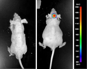
Figure: Expression of luciferase in U87-Luc MG orthotopic model. Control (left) and implanted glioma mouse model fluorescence (right) was captured after intraperitoneal luciferin injection (10 min incubation).
View details of the case study here.
Get Instant Quote for
BxPC-3 Xenograft Model
What we offer?
Our in vivo xenograft service department evaluates the efficacy of preclinical and clinical cancer therapeutics utilizing more than 50 validated immunocompromised xenograft mouse models. The value of utilizing our xenograft service department is highlighted by the ability to completely characterize the efficacy, dose regimen, dose levels and optimal combination ratios of lead compounds for cancer, obesity, diabetes, infections and immunology research.
During the design and execution of the xenograft study, our scientists will communicate with and assist the client’s decisions regarding these details:
- Study Group Formation: classification of mice by body weight, tumor size or other parameters
- Cancer Cell Line: use of in-house cell lines or utilization of customer-provided cell lines
- Tumor Implantation: intraperitoneal, subcutaneous, submuscular or intravenous
- Test Compound Administration: intraperitoneal, intravenous, tail vein, subcutaneous, topical, oral gavage, osmotic pumps or subcutaneous drug pellets
- Sample Collection: Tumors/tissues can be fixed in 10% NBF, frozen in liquid N2 or stabilized in RNAlater; blood chemistry analysis can be performed throughout the in-life portion of study
Vivarium
Our vivarium is designed such that it enables cost-effective and first-rate preclinical effectiveness testing services. All animal handling and maintenance is regulated following IACUC guidelines. Our facility consists of the following:
- IACUC-regulated and GLP-compliant
- Controlled, limited access lab areas
- Disposable cages
- Sterile food and water
- SPF (specific pathogen-free) animals to guarantee pathogens do not interfere with the experiment
- Established animal handling and micro-injection equipment systems, including an animal health observation program
- All studies follow pre-approved SOPs
Our staff understands that each proposed study design is unique and customized to the client’s needs. We also recognize the importance of the delivered results as being confidential, highly reproducible and that 100% of the intellectual property (IP) is owned by the client.
In order to receive a quote for your xenograft study, email us the specific details listed below in order to efficiently begin the study quote process:
- Cancer cell line(s) used in the study
- Number (n=) of animals in each study group
- Number of study groups and control groups
- Tumor implantation route
- Administration route of test compound
- Species of immunocompromised mouse (e.g. NOD/SCID, athymic Nude)
- Treatment and dose schedule
- Study endpoint and analysis (e.g. tumor growth delay, PK/PD, survival, toxicity, drug combinations)
- Samples collected: tumor and tissues to be collected, including storage condition (e.g. snap frozen, RNAlater, 10% NBF, nucleic acid isolation)